Monocyte subsets monitoring – TiMaScan™

Description
Last update: March 20th, 2020
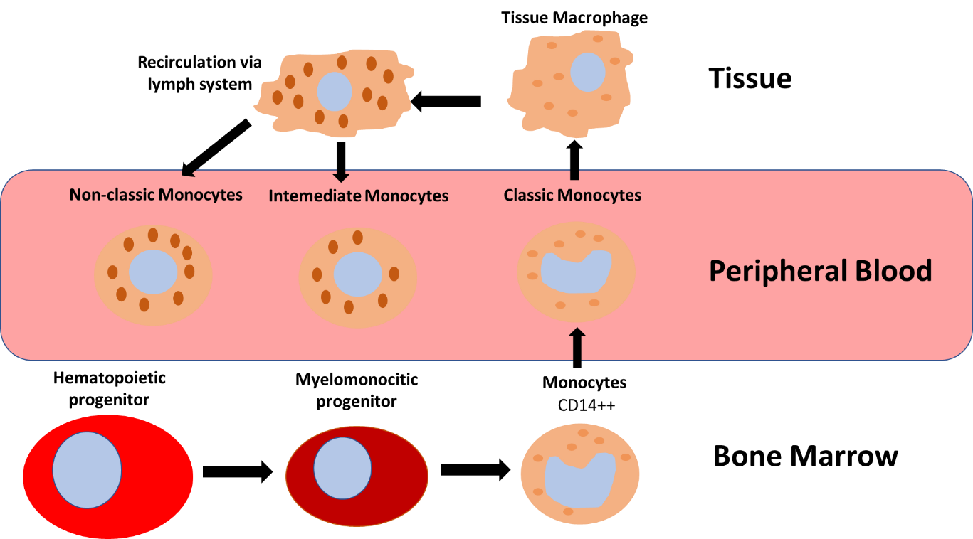
Monocytes are innate immune cells derived from myeloid bone marrow precursors. They circulate in peripheral blood (PB) until they are recruited to clear dying cells and differentiate into tissue macrophages, and are then recirculated.
There are three immunophenotypically different monocyte maturation stages that circulate and can be encountered in peripheral blood:
- Classical monocytes (CD14+CD16-): approximately 80% of all monocytes.
- Intermediate monocytes (CD14+CD16+).
- Non-classical monocytes (CD14-/dim CD16+/++).

Several studies have identified relevant changes in relative and absolute numbers of circulating monocytes/macrophages in the aftermath of several clinical settings, specially involving tissue damage. For this reason, peripheral blood monitoring represents a sensitive test for the detection and quantification of the different subsets of monocytes.
Resources
Publications:
- van der Bossche WBL, et al. Flow cytometric assessment of leukocyte kinetics for the monitoring of tissue damage. Clinical Immunology. 2018 Dec; 197:224-30. Go to publication.
- Damasceno D, et al. Distribution of subsets of blood monocytic cells throughout life. 2019 Jul; 144(1):320-3.e6. Go to publication.
- Kapellos TS, et al. Human Monocyte Subsets and Phenotypes in Major Chronic Inflammatory Diseases. Frontiers in Immunology. 2019 Aug. 10:2035. Go to publication
- Talati, T, et all. Monocyte subset analysis accurately distinguishes CMML from MDS and is associated with a favorable MDS prognosis. Blood. 2017 Mar. 129(13): 1881-3. Go to publication
- Sampath P, et al. Monocyte Subsets: Phenotypes and Function in Tuberculosis Infection. Frontiers in Immunology. 2018 Jul. 9:1726. Go to publication

