Flow Cytometry
The flow cytometer
Last update: October 1st, 2019
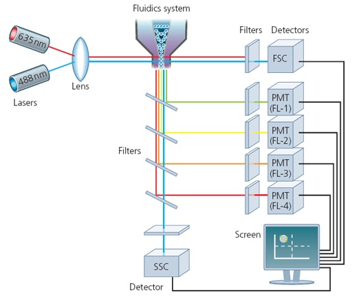
Flow cytometry uses fluorescent probes to identify and characterize cells or particles. Cells or particles tagged with fluorescent molecules enter the cytometer via a fluid stream. The cells then pass by a laser, which emits a specific wavelength of light. The fluorescent probes are excited by the laser and then emit light. The scattered light (intrinsic parameters that evaluate size and complexity), and fluorescence emissions of each particle, are detected and amplified, then translated into an electronic signal, which is sent to the computer where the distribution of the population with respect to the different parameters is represented. The result is a visual presentation describing an individual or group of cellular events.
There are 3 main components of a flow cytometer—the fluidics, optics and electronics—that work together to provide a complete system of cell analysis:

Fluidics system: it is responsible for transporting sample from the sample tube to the flow cell. Once through the flow cell (and past the laser), the sample is either sorted (in the case of cell sorters) or transported to waste.
Optical system: its components include excitation light sources, lenses, and filters used to collect and move light around the instrument and the detection system that generates the photocurrent.
Electronics system: here, the photocurrent from the detector is digitized and processed to be saved for subsequent analysis.
Today’s instruments offer an increased number of detectable fluorescent parameters (from 1 or 2 up to ~30 or more), all measured at the same time on the same cell. Because of its speed and ability to scrutinize at the single-cell level, flow cytometry offers the cell biologist the statistical power to rapidly analyze and characterize millions of cells, albeit at the expense of the morphological characteristics and subcellular localization that microscopy can provide.
Resources
Publications:
- Adan A, et al. Flow cytometry: basic principles and applications. Crit Rev Biotechnol. 2017 Mar;37(2):163-176. Go to publication
- Cossarizza A, et al. Guidelines for the use of flow cytometry and cell sorting in immunological studies. Eur J Immunol. 2017 Oct;47(10):1584-1797. Go to publication
- Piyasena ME, et al. Multinode acoustic focusing for parallel flow cytometry. Anal Chem. 2012 Feb; 84(4): 1831–1839. Go to publication
- Ward M, et al. Fundamentals of Acoustic Cytometry. Curr. Protoc. Cytom. 49:1.22.1-1.22.12. Go to publication
- Brown M and Wittwer C. Flow Cytometry: Principles and Clinical Applications in Hematology. Clinical Chemistry. 2000 April;46:8(B)1221–1229. Go to publication
- Howard M. Shapiro Practical flow cytometry, 4th ed. John Wiley hi Sons, Inc. 2003 August; 736 Pages. Go to publication

